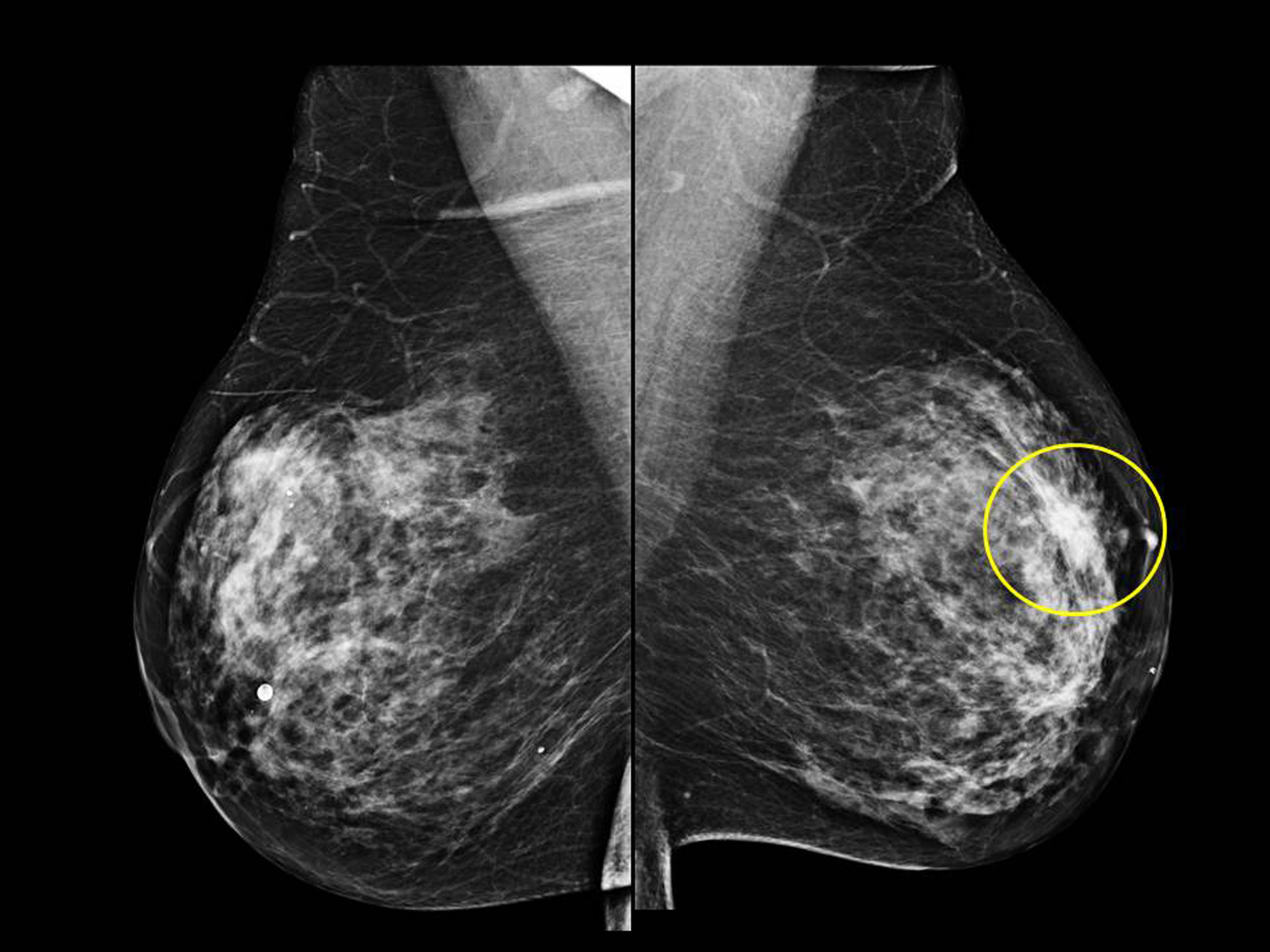
What Does Breast Cancer Look Like On A 3D Mammogram / 3D Mammograms | Beverly Hills | Bedford Breast Center - The tumor cells don't stay within the clear borders of the mass, but instead invade the nearby breast tissue.
Dapatkan link
Facebook
X
Pinterest
Email
Aplikasi Lainnya
What Does Breast Cancer Look Like On A 3D Mammogram / 3D Mammograms | Beverly Hills | Bedford Breast Center - The tumor cells don't stay within the clear borders of the mass, but instead invade the nearby breast tissue.. Images are displayed as a series of thin slices that can. In the mammogram below, one can see the increase in the density of the fibroglandular tissues behind the nipple. American cancer society, 9 oct 2017. Essentially, mammograms turn a 3d object into a 2d object. Finding breast lumps and seeing change in the size and shape.
These deposits show up as tiny white spots on a mammogram, and there may be only one or two, or too many to count, says jay baker, md, a breast imaging specialist at the duke cancer center. Digital breast tomosynthesis (tomo), also known as 3d mammography, is a revolutionary new screening and diagnostic breast imaging tool to improve the early detection of breast cancer. However, when the breast is compressed from top to bottom, the tissue in the upper breast can overlap tissue in the lower breast. Several patterns of calcifications are seen with dcis, including: Microcalcifications, which look like white specks on a mammogram.
Do You Need a 3-D Mammogram? | SafeBee from media.safebee.com A tumor that is benign, it is not a health problem and it may not grow or change shape. Several patterns of calcifications are seen with dcis, including: Finding breast lumps and seeing change in the size and shape. The outer edges of these cells look fuzzy or spiky (called spiculated). The tumor cells don't stay within the clear borders of the mass, but instead invade the nearby breast tissue. Calcifications are calcium deposits within the breast tissue and they look like small white spots. A false positive is when a mammogram shows an abnormal area that looks like a cancer but turns out to be normal. This decreases the overlap and makes it easier to see cancers. additionally, the combination of 2d and 3d mammogram imaging has been shown to reduce false positives that require a patient to return to the clinic for additional screening.
They are often caused by aging, an old injury, or inflammation and are usually benign.
If found in an area of rapidly dividing cells or grouped together in a certain way, they may be a sign of dcis or breast. Bright spots on a mammogram that look like potential tumors could turn out to be overlapping tissues or a blood vessel folding over on itself, friedewald said. The doctor reading your mammogram will be looking for different types of breast changes, such as small white spots called calcifications, larger abnormal areas called masses, and other suspicious areas that could be signs of cancer. However, when the breast is compressed from top to bottom, the tissue in the upper breast can overlap tissue in the lower breast. We'll show you breast cancer pictures to help you identify any physical traits of the condition. Digital breast tomosynthesis (tomo), also known as 3d mammography, is a revolutionary new screening and diagnostic breast imaging tool to improve the early detection of breast cancer. However, in rare cases, breast cancer can be the cause of gynecomastia so, a full mammographic investigation is always necessary. Microcalcifications, which look like white specks on a mammogram. That makes it easy to detect abnormalities, which generally show up as white. The look of breast cancer on a mammogram a tumor or lump will appear as a focused white area on the mammogram. Dense breast tissue appears solid. Screening mammograms have been used since the 1980s. A 3d mammogram is used to look for breast cancer in people who have no signs or symptoms.
If a doctor suspects an abnormal growth in the breast after a clinical breast examination or screening mammography, evaluation of breast cancer ultrasound images will help confirm the diagnosis. A diagnostic mammogram is used to check for breast cancer when there is a sign or symptom of disease. This overlapping tissue can cause the resulting image to look like cancer. The tumor cells don't stay within the clear borders of the mass, but instead invade the nearby breast tissue. 1 the gray areas correspond to normal fatty tissue, while the white areas are normal breast tissue with ducts and lobes.
RSNA 2013: Breast Cancer Prognosis Potentially Affected by ... from healthmanagement.org One advantage of ultrasound technology is that it allows substantial freedom in obtaining breast images from any orientation. This decreases the overlap and makes it easier to see cancers. additionally, the combination of 2d and 3d mammogram imaging has been shown to reduce false positives that require a patient to return to the clinic for additional screening. Essentially, mammograms turn a 3d object into a 2d object. Digital breast tomosynthesis (tomo), also known as 3d mammography, is a revolutionary new screening and diagnostic breast imaging tool to improve the early detection of breast cancer. What does breast cancer look like? To license this video for patient education or content marketing, visit: A 3d mammogram is used to look for breast cancer in people who have no signs or symptoms. The outer edges of these cells look fuzzy or spiky (called spiculated).
Normal breast tissue can look 100,000 different ways on a mammogram.
What does breast cancer look like? If a doctor suspects an abnormal growth in the breast after a clinical breast examination or screening mammography, evaluation of breast cancer ultrasound images will help confirm the diagnosis. Macrocalcifications, which look like small white dots on a mammogram. A 3d mammogram is used to look for breast cancer in people who have no signs or symptoms. Breast cancer can appear as a spiculated mass, cluster of tiny calcifications, smoothly marginated mass, area of subtle distortion or be invisible on. This decreases the overlap and makes it easier to see cancers. additionally, the combination of 2d and 3d mammogram imaging has been shown to reduce false positives that require a patient to return to the clinic for additional screening. What does breast cancer look like on a mammogram? Finding breast lumps and seeing change in the size and shape. The most common presentation of dcis on mammography involves the appearance of calcifications. A screening mammogram is performed at regular intervals to check for breast cancer in women who have no signs or symptoms of the disease. Bright spots on a mammogram that look like potential tumors could turn out to be overlapping tissues or a blood vessel folding over on itself, friedewald said. That makes it easy to detect abnormalities, which generally show up as white. Diagnostic mammograms involve taking more views than screening mammograms.
Diagnostic mammograms involve taking more views than screening mammograms. What does breast cancer look like? A mammogram can show breast changes such as calcifications, masses, or other symptoms that might be cancer. The outer edges of these cells look fuzzy or spiky (called spiculated). We'll show you breast cancer pictures to help you identify any physical traits of the condition.
Cancer Chronicles: Mammogram and Ultrasound from 1.bp.blogspot.com One advantage of ultrasound technology is that it allows substantial freedom in obtaining breast images from any orientation. Macrocalcifications, which look like small white dots on a mammogram. That's only half the story.. If a breast radiologist can make that mistake, anybody can make that mistake. even if patient x weren't the mother of a close friend, her case made the radiologist reconsider the impact skin markers have in mammography, even with digital breast tomosynthesis. What does breast cancer look like? If a doctor suspects an abnormal growth in the breast after a clinical breast examination or screening mammography, evaluation of breast cancer ultrasound images will help confirm the diagnosis. A number of studies have found that 3d mammograms find more cancers than traditional 2d mammograms and also reduce the number of false positives. However, when the breast is compressed from top to bottom, the tissue in the upper breast can overlap tissue in the lower breast.
Screening mammograms have been used since the 1980s.
It appears to be developing in a concentric pattern. A diagnostic mammogram is used to check for breast cancer when there is a sign or symptom of disease. This is why you should always talk to your doctor if you notice an unexplained change in the size of a breast. The most common presentation of dcis on mammography involves the appearance of calcifications. If a doctor suspects an abnormal growth in the breast after a clinical breast examination or screening mammography, evaluation of breast cancer ultrasound images will help confirm the diagnosis. One advantage of ultrasound technology is that it allows substantial freedom in obtaining breast images from any orientation. Moose & doc breast cancer, 21 may 2018. This decreases the overlap and makes it easier to see cancers. additionally, the combination of 2d and 3d mammogram imaging has been shown to reduce false positives that require a patient to return to the clinic for additional screening. However, 3d allows us to 'slice through' the breast, making thin sections, like on a ct scan. Any area that does not look like normal tissue is a possible cause for concern. That makes it easy to detect abnormalities, which generally show up as white. A 3d mammogram is used to look for breast cancer in people who have no signs or symptoms. Macrocalcifications, which look like small white dots on a mammogram.
Daftar lengkap pilih bidang usaha klasifikasi baku lapangan usaha indonesia. Khasiatnya untuk penyakit di kepala, sinus dan masalah2 mata. 31.10.2021 · "kita tidak tahu kapan pandemi selesai. Sebagai tanda terima kasih, maka dr. Menyembuhkan sultan kutai yang telah lama jatuh sakit. dr. Ida Bagus Surya Putra Manuaba, M.Kes.Sp.THT-KL.MARS from baliroyalhospital.co.id Kategori (a s/d u), golongan pokok (2 digit), golongan (3 digit) dan kelompok (5 digit). Pt bank syariah indonesia tbk. Menyembuhkan sultan kutai yang telah lama jatuh sakit. Niken ageng rizki obsesitas dapat berpengaruh pada lingkar leher dan jalan napas, sehingga membuat seseorang mendengkur. Menteri tenaga kerja dan transmigrasi. Sebagai tanda terima kasih, maka dr. Menyembuhkan sultan kutai yang telah lama jatuh sakit. Sebagai tanda terima kasih, maka dr. ...
18.01.2021 · 30 best sad movies on netflix to stream when you want a good cry. Read on for our picks of the saddest movies on netflix, and for more ways to pass the time, check out these 16 netflix shows you can watch from start to finish this weekend. Here's everything new on netflix in august 2021 catch me if you can From historical drama to heartbreaking romance, there's every kind of tearjerker. 21.09.2021 · netflix wants to make animated and live action films and tv, publishing, games, immersive experiences, live theatre, consumer products and more. by j. Pin On Entertainment from i.pinimg.com 16.04.2021 · there's something for everyone, as long as you've got tissues by your side. 21.09.2021 · netflix wants to make animated and live action films and tv, publishing, games, immersive experiences, live theatre, consume...


Komentar
Posting Komentar